Blog
Dental hygiene tips for healthy teeth & gums
Panoramic Dental X-Ray: What It Is, How It Works, and What It Shows
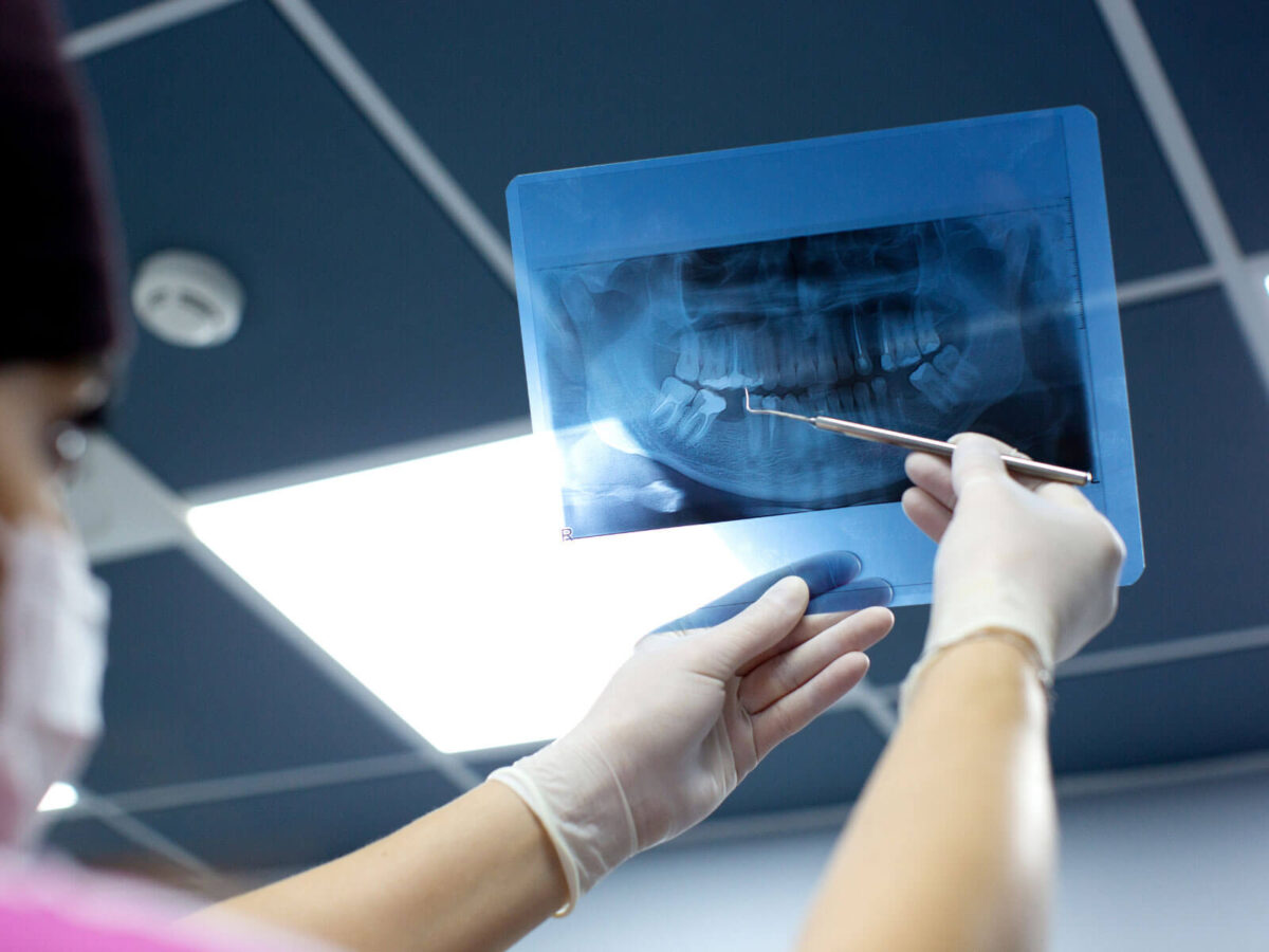
A dentist glances at a screen and says “impacted” before explaining anything further. Whatever image they’re looking at usually isn’t the small bite-down film most people picture, the kind that only shows two or three teeth at a time. It shows both jaws, every tooth, laid out flat in one wide shot, sinuses and jawbone included.
That’s a panoramic dental X-ray, and it looks nothing like the small film most people expect. Instead of two or three teeth at a time, the whole mouth shows up in one wide image, jaws, sinuses, and all.
What a Panoramic Dental X-Ray Actually Is
There’s no biting down on film here. Cleveland Clinic files this under extraoral imaging, meaning the sensor stays outside the mouth instead of being pressed against the teeth.
One flat image comes out the other end, both arches side by side, every tooth included, sinuses above, jaw joints near the ears. Dentists sometimes call it an orthopantomogram, though almost nobody outside the office uses that word. “Pano” sticks, or just a panoramic dental X-ray on the paperwork.
How the Machine Physically Captures the Image
The setup looks strange the first time someone sees it. An arm extends from a column, X-ray source on one end, moving sensor on the other, head positioned in between.
It rotates slowly, tracing something close to the shape of the jaw, ten to twenty seconds total, though the beam only hits any single point for a sliver of that time. A chin rest and forehead guide hold everything steady. Move mid-scan, and the image blurs, often enough that the whole thing has to be redone.
Film used to mean waiting for it to develop. Digital sensors replaced that in most offices years ago, and the exposure dropped along with it, roughly 80 to 90 percent lower, according to the Cleveland Clinic. That’s a big part of why a panoramic dental X-ray isn’t reserved for unusual cases anymore.
What Shows Up on the Image
Everything inside that sweep gets flattened into one picture, both arches, roots, and surrounding bone. The maxillary sinuses sit as darker patches above the upper teeth. The TMJs show up near the back on both sides.
Wisdom teeth still buried under the gum are the classic catch here, especially ones angled sideways into a neighbor instead of coming in straight. Teeth that never erupted at all show the same way, sometimes spotted years before symptoms would’ve flagged anything.
Cysts and tumors get picked up too, and this is where the width earns its keep. These grow slowly, painlessly, tucked into bone, a mirror and a light would never reach. On a pano, they show up as a patch that looks off, dense, or dark in a spot that shouldn’t be either.
Implant planning leans on one specific detail: where the nerve running through the lower jaw actually sits. Get an implant post too close, and the result can be lasting numbness in the lip or chin. A panoramic dental X-ray maps that nerve’s rough path before anyone picks up a drill.
What This X-Ray Tends to Miss
Width costs some detail. Flatten a curved jaw into one image, and the center stays sharp while everything at the edges softens, the same way a wide photo loses crispness away from the middle.
Small cavities between two teeth are the clearest casualty. Bitewing X-rays exist specifically for that gap. The ADA’s own overview of dental imaging, run in JADA, points to bitewing and periapical films as the standard for catching decay and root-level changes tooth by tooth, a level of resolution this wider scan was never built to deliver.
Most dentists don’t pick one over the other. They use both. A panoramic dental X-ray answers whether anything’s structurally off across the whole jaw. Bitewings answer whether one specific tooth is starting to decay. Skip one, and there’s a real gap, depending on what’s being checked.
Who Actually Ends Up Getting One
New patients, often. No history to compare against yet, so a single wide image gives a starting point fast instead of piecing one together from several narrow shots. MouthHealthy notes dentists sometimes order X-rays for new patients specifically to set that baseline and catch anything that shifts later.
Orthodontic planning needs it too. Fitting braces means seeing how every tooth lines up against the whole jaw, not just how a few of them look up close. Teenagers get it a lot for wisdom tooth tracking, watching third molars that haven’t fully come in yet.
Implants, dentures, and unexplained jaw pain all point back to the structure under the visible teeth rather than the teeth themselves, which is exactly what a panoramic dental X-ray is built to show.
None of that means it happens at every visit. ADA guidance from 2024 leaves the call to clinical judgment for that specific patient, not a fixed schedule everyone gets slotted into regardless of age or history.
How Often Does This X-Ray Actually Need Repeating
There’s no fixed clock here, and that surprises people. There’s no universal “every X years” rule stamped on a panoramic X-ray the way there sort of is for bitewings.
The ADA’s newest recommendations, published jointly with the American Academy of Oral and Maxillofacial Radiology, confirm dental X-rays should only be ordered when clinically necessary, which puts the decision back on individual risk factors rather than a calendar.
Age moves the needle more than almost anything else. A teenager still growing wisdom teeth might need one every year or two just to track eruption. An adult with a stable, uncomplicated mouth might go five years or longer without needing another one at all.
Certain events reset the clock regardless of age. Planning an implant, evaluating unexplained jaw pain, or investigating a lump that showed up somewhere in the mouth all tend to trigger a new panoramic dental X-ray on the spot, timeline aside.
The ADA frames this as following the ALARA principle, as low as reasonably achievable, meaning the goal is getting the image only when it’ll change something about the treatment plan, not repeating it out of habit.
What Happens During The Visit
Setup takes a bit longer than the scan itself. A technician adjusts the chin rest, has the patient bite gently on a small plastic tab, and checks alignment before starting anything.
A lead apron often goes on first. Cleveland Clinic mentions a thyroid collar often gets added too, mostly as extra caution given how small the dose already is.
The arm moves next, a low hum, maybe a faint click. Nothing touches skin, nothing gets injected, nothing goes inside the mouth beyond that bite tab. Digital equipment means the image is usually up on a monitor within a minute, the dentist already looking over the panoramic dental X-ray before the patient’s even out of the chair.
FAQ
Are panoramic dental X-rays hurtful?
No. No needle, no drilling. Biting lightly on a small tab while the arm circles around is the entire physical sensation involved.
Why this instead of the usual small X-rays?
Depends what’s being checked. Wisdom teeth, implant planning, jaw pain, anything involving the whole jaw structure tends to call for the wider image instead of a close-up of a few teeth.
More radiation than a regular dental X-ray?
It’s comparable, sometimes less. Digital panoramic equipment uses a fraction of what older film machines needed.
Can it catch cavities?
Larger ones, usually. Small ones between teeth tend to slip past it, which is exactly what bitewings are for instead.
Do kids get these too?
Frequently, mainly to track how permanent teeth are developing under the gum and whether there’s room for everything to come in.
Conclusion
A panoramic dental X-ray isn’t a bigger version of the small films most people know. It’s a different tool, built for the whole jaw at once instead of a handful of teeth in fine detail. That’s how it catches an impacted wisdom tooth or a jaw cyst a bitewing would never see.
Implants, orthodontics, and wisdom teeth coming in are all common reasons this scan comes up. Worth asking directly what it’s looking for in that specific case, and worth booking an appointment if it’s been a while since the jaw itself, not just the teeth, has been checked.